
Get full access with a free account
Benefits of the Coloplast® Professional Educational platform
- Full access to educational content, events and resources
- Track your progress
- Share content with your colleagues
- Share supporting material with your patient
80% of non-healing wounds contain biofilms1,2
- A biofilm is a structured community of microorganisms with genetic diversity that creates chronic infection2
- Biofilms are characterised by significant tolerance to antibiotics and antimicrobials while remaining protected from host defense2
Biofilms are not visible to the eye, but signs of infection indicate presence of biofilms, e.g.:
- Delayed wound healing
- Increased exudate and slough
- Increased inflammatory response
- Erythema
- Malodour
- High bacterial load
Biofilm, or bacterial aggregates, are microorganisms embedded in a thick, slimly barrier of sugars and proteins. This barrier shields microorganisms from a patient’s natural immune system and from many antimicrobial agents.1 Biofilm can form in a wound within 24 hours.
Bacteria are often viewed as being single cells that multiply rapidly when in exponential growth. This is referred to as ‘planktonic form’ and relates mostly to acute infections. Bacteria can also form aggregates, or communities, of slow-growing cells – this kind of formation is referred to as ‘biofilm’.
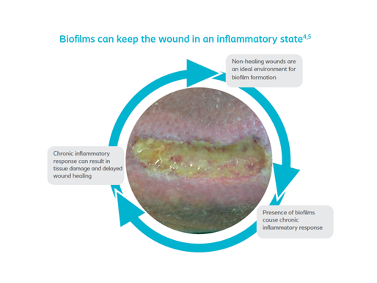
How do biofilm affect wound healing?
Biofilm are probably the most important single cause of persistent, delayed healing in wounds.2 As figure 1 illustrates, they are also thought to delay wound healing by effecting an inappropriate inflammatory response, which is ineffective, poorly orchestrated and damaging to host tissues.2
There is increasing evidence that biofilm are present in most, if not all, chronic, non-healing wounds.3 Therefore, if you diagnose that your patient has an infection in a chronic wound, it’s recommended that you follow the guidelines for preventing and managing biofilm.2
Two types of bacteria formations
The most common biofilm formers are Staphylococcus aureus and Pseudomonas aeruginosa.2
S.aureus, a Gram-positive bacteria, that:
- is a prominent human pathogen
- a keen biofilm former
- ranges from minor self-limited skin to invasive life-threating infections
30% of all healthy individuals carry S. aureus in the nose without any awareness or impact.1
A very well-known type of S. aureus is MRSA with continuing mortality rates of 28-38%. 4
- Pseudomonas aeruginosa is a Gram-negative microorganism that is believed to reside in the deep wound tissue, and is a keen biofilm former.1
- 52.2 % of chronic leg ulcers contained P. aeruginosa and those wounds are characterised by larger sizes and slower healing rates. 1
- P. aeruginosa produces pigments, which may aid in visual detection of a P. aeruginosa, through green discolouration.2
Example of wound biofilm
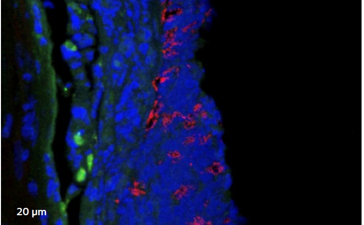
Confocal laser scanning microscopy (CLSM)
Image 1 shows a microscopic image of biofilm (highlighted in red), with clusters often less than 1/10 mm. This results in many swabs coming back inconclusive.
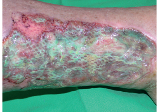
Example of wound with suspected biofilm
You should suspect biofilm in ‘healable’ wounds, that are non-healing, if you have taken all of the appropriate measures.1
How do you detect biofilm?
Detecting biofilm can be challenging, due to the following factors:
- Biofilm are microscopic structures, which are invisible to the naked eye. So you need to use a high-powered microscope to detect them.
- In a clinical setting, the best detection method is a tissue biopsy. However, biofilm are small and unequally distributed in the wound bed. A wound may have different species of biofilm, and they are typically scattered around in small, isolated, single-species islands. That’s why they can be easy to miss.
When should you suspect biofilm?
Try asking yourself the following questions2:
- Have all appropriate diagnostic and therapeutic measures been followed?
- Is the wound failing to heal as expected?
- Does the wound show signs of local infection or inflammation?
If you answer ‘Yes’ to at least 2 of these questions, it would be clinically relevant to treat for biofilm.